Featured

Why I love what I do?

I wasn’t a natural at being a doctor and struggled a little bit in medical school. Medical school was sometimes confusing and intimidating. I picked up empathy along the way and learnt to be compassionate to other people. I found out that I loved the difference that I bought to others and got better at doing my job. A lot of what I love about my job today, I discovered while being a doctor. If I hadn’t taken up medicine I would have never found out how much I would enjoy it.

Once I started residency, I took to Ophthalmology like a fish to water. It seemed to tick off all the right boxes for me. Ophthalmology is an awesome subject and it had the good mix of medical and surgical management. As I got deeper into the subject, the more I began loving it. I discovered that surgery gave me the high I had never experienced before in my life which prompted me to try to become a better surgeon every day. I learnt that there is a lot of suffering between life and death- and there was something I could do about it. I think I did well in it and in time my surgical prowess improved too. I had wonderful teachers who provided me the best platform to be a skilled surgeon that made me so confident that I always carried a chip on my shoulders-maybe even now!

I found an interest in a niche sub-specialty called Ophthalmic Plastic Surgery. The tipping point of my residency when I decided for sure that Oculoplasty was my calling was when I assisted my boss in a surgery called- Lateral orbitotomy-where he was removing a tumour from the back of the eye cutting through bone. While delivering the tumour out, I said to myself then, “that is something I want to do for a living!” . To cut a long story short I eventually managed to get into a fellowship that fed into my obsession. It was a coveted fellowship and my dream come true. I learnt to be pretty good at Oculoplasty- mostly because of my excellent training by my mentors in fellowship and equally too because I loved what I did so much.

Then I got back to Bangalore to set up my own exclusive practice in Ophthalmic Plastic Surgery and Ophthalmic Oncology. All the training till then had not taught me the lessons I learnt while establishing my own practice. It was tough starting out and I did struggle for the first couple of years. It is a lot better now and there is definitely a fair distance still to travel. I’m happy however that I found my calling in the midst of it all even though I didn’t have any grand plan all along.

My teachers in medical school, internship, residency and fellowship taught me more than just medicine & surgery- How to deliver bad news, how to hold a patient’s hand while he or she is passing through a difficult phase of treatment, how to negotiate with an obstinate non-compliant patient, how to know where you can fit into the patient’s convalescence and when to get out- these are not written in textbooks but learnt by spending time training with my role models. Over the years during my training and later in practice I would see time and again that the doctors whom I wanted to emulate owned their patients. I hope I did that over time.

Every day, I get up in the morning hoping that my day is filled with cases which pique my curiosity. In my clinics and in the operating theatre, I’m never disappointed- I get presented with fresh challenges constantly which always keeps me on my toes. Whether fixing a facial fracture or removing a large tumour behind the eye or making someone look a better version of themselves, life is never boring for me. What my work gives me is more than I can describe in mere words but I guess these above paragraphs will have to do for now.

It hasn’t been easy. Like I said- I wasn’t a natural. I have faltered many times and suffered self-doubt more times than I can count. This “successful” journey has been inundated with many failures along the way which makes me cherish what I have even more. In all this, I have been lucky to have had teachers and mentors who tapped into my potential and sometimes saw more in me than I saw myself. My family- my parents and my wife- have stood behind me like a rock-without whose constant support I wouldn’t have been able to take the crazy career decisions I did. Despite their reservations, they trusted my madness and never once told me to hold back and “settle down”.

Last but not the least-my patients. I’m lucky to be born in a country which is so diverse as is challenging and in an era where I as a medical professional can make so much difference using the latest in my field. I often crib about my most difficult patients-but not today. My patients have brought me more joy than grief and I have many times been touched by their kindness and overwhelmed by their gratitude. However, they don’t realise that I get paid to do what I love most and I owe them more than they owe me!

How do you correct hereditary acquired droopy eyelids?

This 75yr old was an avid cyclist🚴‍♀️ , swimmer 🏊‍♀️ and Yoga 🧘 practitioner. He has been severely restricted from regular activities due to his bilateral ptosis. He was suffering from Chronic Progressive External Opthalmoplegia (CPEO). It is a genetic condition that affects patients mainly in the second half of their lives. His two elder brothers too suffered ptosis without getting it corrected and passed on without relief. 

Chronic progressive external ophthalmoplegia (CPEO) describes an spectrum of hereditary myopathies affecting extraocular muscles (EOMs)   commonly manifesting as bilateral ptosis and near total external ophthalmoplegia. 

As the name suggests, it is a chronic, progressive, bilateral, typically symmetric, and external (i.e., spares the pupil) ophthalmoplegia. CPEO is associated with mitochondrial disease and can occur as isolated oculomotor symptoms (isolated CPEO) or in conjunction with other systemic findings (“CPEO-plus”). Depending on additional clinical features, CPEO can be evaluated as part of a systemic myopathic or neurologic syndrome

Surgical correction of Myogenic Ptosis like in CPEO is challenging as conventional methods cannot tackle this condition safely as both opening and closure of eyelids is compromised in this condition. 

However, this gentleman proved to be luckier than his elder brothers. When he approached me, I told him about the technique I was experimenting with which was having acceptable results- Tarsal Switch technique for correction of severe myogenic ptosis. He agreed to go ahead despite a detailed counselling mentioning that there might be chances it may not work in everyone who has CPEO. He told me, “he had nothing to lose, even if I have a 50% chance I’ll accept it”. 

Post surgery, the surgery was a success-as can be seen by the photo collage above. We were able to achieve clearance of the central visual field. The patient is thrilled at the results and is back to his previous active lifestyle 🧘🚴‍♀️🏊‍♂️.

My experience over the past 2+ years with this technique has been gratifying and I have made some modifications to the techniques described in the literature. I’m surprised why this technique is not being offered more frequently. 

⏲Surgery time: 45mins per side
💉Anaesthesia: Done under Local with or without sedation
🗓️Downtime: 3-4 weeks
📈Duration of effect: Long term 
📠 Phone: +91 80 2502 3257
📩 E-mail: raghuraj@drraghurajhegde.com
🖥️ Website: www.drraghurajhegde.com

©All patient photos are being used with the express consent of the patient. These cannot be shared or reproduced elsewhere.

Customised Implants : Precision Surgery

Patient Specific Implant (PSI) have revolutionised the treatment of secondary orbital reconstructions. The above picture red broken line shows level of the eye balls before and after secondary reconstruction of left orbit with a PSI. 

Picture showing the before and after surgery pictures of a male patient after correction of eye socket deformity with a patient specific implant.

This young man suffered from a Road traffic accident about a year back before he come to see us. He had a left orbital floor fracture with bilateral inferior orbital rim fracture at the time of injury. He refused surgery at the time. Below you can see the old and new 3D reconstruction CT scans dated more than a year apart.

The old and new 3D reconstruction CT scans dated more than a year apart.

However, slowly he started developing Left eye enophthalmos (eye sinking into eye socket), left orbital deformity compared to right eye. In addition, he had restriction in movement for upgaze and diplopia (double vision) in certain gazes. He was advised a PSI for correction of this late deformity. 

Virtual planning: 
🖥️⌨️💻👩‍💻🧑‍💻💽🖱️

A PSI is designed using the normal side mirrored to the other side as a template. The customisation and design modifications is done by collaboration of the operating surgeon 👨‍⚕️with the bio-medical design engineers 👩‍💻via video-conferencing calls. 

The resulting design is used for  3D printing the implant mock up and the final implant on approval of the mock implant. Below you can see the mock up as well as 3 D printed orbital implants 👀 on the 3D printed anatomical skull model 💀 of the patient made of plastic. 

The mock up as well as 3 D printed orbital implants on the 3D printed anatomical skull model of the patient made of plastic. 

His post operative CT scan shows perfect correction of his Floor fracture and zygoma augmentation.

In addition to correcting the facial deformity, both enophthalmos and diplopia on upgaze was completely resolved. Needless to say, the patient was thrilled. 

Implants can be made up of Titanium (commonly), Polycaprolactone (Bioresorbable/Osteo-integrative implant) or the newest kid on the block Poly Ether Ether Ketone (PEEK). PEEK is a semi-crystalline linear polycyclic thermoplastic that has been proposed as a substitute for metals in orbital implants.

PSI is an excellent modality to provide precise, predictable and reproducible results resulting in both aesthetic and functionally good outcomes. 

📆Virtual Planning and PSI 3D printing time: 2 weeks
⏲Surgery time: 2 hours 
💉Anaesthesia: Done under general anaesthesia 
🗓️Downtime post surgery: 3-4 weeks
📈Duration of effect: Long term 
📠 Phone: +91 80 2502 3257
📩 E-mail: raghuraj@drraghurajhegde.com
🖥️ Website: www.drraghurajhegde.com

©All patient photos are being used with the express consent of the patient. These cannot be shared or reproduced elsewhere

It seems like Magic but it’s NOT!

Ocular Surface Squamous Neoplasia (OSSN) is a spectrum of pre-cancerous and cancerous lesions of the conjunctival epithelium (transparent coat covering the surface of the eye).

This condition can be diagnosed clinically and if promptly treated can achieve complete cure-as in this case. The before and after photo of the patient shows that the lesion is completely removed and looks like there’s no surgery at all. If not operated on time, the cancer will invade into the eye can cause vision loss and can even become fatal.

Below is the picture of the lesion at presentation and with lissamine green staining. The keratin deposits and lissamine green staining on the lesion with feeder vessels leading up the lesion is pathognomic of OSSN.

Below is the before after picture in static mode so that you can appreciate the difference. 

⏲Surgery time: 30-45 mins
💉Anaesthesia: Done under Local
🗓️Downtime: 3-4 weeks
📈Duration of effect: Long term 
📠 Phone: +91 80 2502 3257
📩 E-mail:dr.raghuraj.hegde@gmail.com
🖥️ Website: www.drraghurajhegde.com

Surgery:
A no touch technique is used to excise the lesion with 4mm margins, alcohol keratectomy to excise involved corneal epithelium-followed by double freeze-thaw cryotherapy of conjunctival edges. The ocular surface reconstruction is done using Conjunctivo-Limbal Auto-Graft (CLAG) and Amniotic Membrane Grafting (AMG) gives best outcomes without complications.

Happy tears!

This sweet 84yr old lady came to us with a painful blind right eye. We advised eye removal (evisceration) with orbital implant followed by customized ocular prosthesis. She had been resisting this surgery the past 5 years when the blind eye of 20yrs started becoming painful. We finally managed to convince her for the surgery and went ahead with the surgery. Our talented ocularist, Mr. Jibran Munnavar put in incredible work in making the customised prosthesis for her post surgery. 

After the prosthesis fitting, when I showed the lady and her daughter the pre-operative photos, the daughter broke down in tears and told me “You’ve given back my mother after 20yrs”. 

Small things we do for our patients mean so much for them personally. This also proves that it is never too late to be yourself again. ❤️

A #Customized ocular prosthesis (prosthetic eye) 👁️ surgery is considered when we have to remove an #eye due to one of the following reasons:
1. Eye Cancer (for example Retinoblastoma in children)
2. Irreparable eye injury due to trauma (children & adults)
3. Cosmetically disfigured eyeball with no hope of visual rehabilitation and which may or may not be painful.

There are two stages in achieving great outcomes like in this case.

Stage 1- #Surgery performed by an #oculoplastic surgeon. The surgery involves #Enucleation (eyeball being removed)/#Eviceration (the contents of the eyeball removed retaining the outer white scleral shell) and globe volume is then replaced with an appropriately sized #orbital #implant.

Stage 2- After the eye socket heals which takes about 6 weeks, an #ocularist (a professional who fabricates prosthetic eyes) then fabricates a customised ocular prosthesis which fits perfectly in the patient’s socket. This prosthetic eye looks similar to the other eye including co-ordinated movements making it look natural. 

Schematic Diagram of ocular prosthesis in relation to orbital implant

For details of this surgery and for appointments

⏲Surgery time: 45 mins
💉Anaesthesia: Done under Local/Sedation
🗓️Downtime: 6 weeks
📈Duration of effect: Long term 
📠 Phone: +91 80 2502 3257
📩 Email:dr.raghuraj.hegde@gmail.com
🖥️ Website: www.drraghurajhegde.com

Eyes don’t see what the mind doesn’t know!

It is not often that an eye surgeon is able to awe a room full of medical students and residents in a busy Ophthalmology clinic. So I’ll take this opportunity to write the story about this this extra-ordinary accomplishment. 

An 18yr old male patient was referred to me from elsewhere. The patient came with complaints of a swelling on his left eyebrow since last 3 months. He had gone to several doctors of different specialties before me and had several provisional diagnosis ranging from Sebaceous Cyst to Schwannoma to Dermoid cyst! 

At first look it did seem to be that one of those diagnoses was right. On examination though, the texture felt like the classical “bag of worms” sign of a particular condition. I asked for history including leading questions of specific signs which would confirm what I suspected. All negative! 

The clincher was by the slit lamp! Lisch Nodules-yellow to brown coloured melanocytic harmartomas- dome shaped projecting out of iris which are harmless and don’t affect vision. However they are present in up to 94% of Neurofibromatosis type 1. 

Lisch Nodules

I repeated my questions about other signs of NF1. 

Café au lait spots-flat brown medium-large skin spots in the body. 6+ spots is strongly indicative of NF1.

Neurofibromas- Small pea-sized bumps on the skin mostly at the back. All denied again!

However, I was confident enough to ask him to lift his shirt so that I could examine his back. I think I heard a gasp of awe behind me from the medical students/interns/residents who were watching. 

Cafe au-lait Spots and Neurofibromas

The café au lait spots and few neurofibomas spread across his back. 

NF1 confirmed. Patient advised for imaging to exclude other features of NF1. Also advised for excision of left brow plexiform neurofibroma with brow-pexy to correct the cosmetic blemish and restore symmetry of the face

Neurofibromatosis Type 1

Will post this patient’s after surgery picture soon!

For Appointments:
📠 Phone: +91 80 2502 3257
📩 Email: dr.raghuraj.hegde@gmail.com
🖥️ Website: www.drraghurajhegde.com

©All patient photos are being used with the express consent of the patient. These cannot be shared or reproduced elsewhere.

Additional Information

Neurofibromatosis is caused by genetic mutations that either are passed on by a parent or sometimes occur spontaneously during pregnancy. 
The specific genes involved depend on the type of neurofibromatosis:

  • NF1. The NF1 gene is located on chromosome 17. This gene produces a protein called neurofibromin that helps regulate cell growth. The mutated gene causes a loss of neurofibromin, which allows cells to grow uncontrolled.
  • NF2. The NF2 gene is located on chromosome 22, and produces a protein called merlin (also called schwannomin), which suppresses tumors. The mutated gene causes a loss of merlin, leading to uncontrolled cell growth.
  • Schwannomatosis. So far, two genes are known to cause schwannomatosis. Mutations of the genes SMARCB1 and LZTR1, which suppress tumors, are associated with this type of neurofibromatosis.

Catching the Moving Lid!

Ocular Myasthenia Gravis (OMG) is one of the conditions that an oculoplastic surgeon can correct without surgery or any invasive intervention. It is a very satisfying to treat a patient of OMG provided the diagnosis is accurate. It has the rare distinction of being both under-diagnosed and over-investigated in different settings. Clinical examination is the cornerstone of a diagnosis of OMG. The various fancy but expensive testing including antibody titres of Acetyl Choline Receptor Antibody (AchRAb) can only corroborate a strong clinical suspicion but is not gospel as only about ~65% of patients are seropositive for AchRAb.

This particular case was especially challenging to diagnose. The child came in with erratic lid positions- sometime ptotic, sometimes eyelid retraction.  The most sensitive clinical test for OMG- Ice-pack test was equivocal in this child of 5 yrs. AchRAb titres were low and she was euthyroid (strong family history of hypothyroidism-both parents).  It was only on the child’s second visit and on re-examination I could elicit the very subtle clinical sign- Cogan’s Lid Twitch Sign. 

Cogan’s lid twitch sign is one of the clinical tests for Myasthenia Gravis. This test is known to have a sensitivity of 75% but a specificity of almost 99% in some controlled studies. 

This sign is elicited by asking the patient to maintain downward gaze for 10 seconds and then ask them to look upward, finally returning to straight gaze. A positive sign is indicated by the definitive twitch of  the upper lid and sometimes overshooting the superior limbus, soon after the patient has returned to straight gaze.

Ice-pack test has been so useful and quite sensitive to test for OMG, that Cogan’s Lid Twitch sign usually take the back seat. Cogan’s sign proved to be the clincher in this case as the other tests were equivocal.

The child was started on oral Pyridostigmine with steroid induction trial after a Neurology consult. Within a couple of weeks the trial was a success. She is now on maintenance therapy with good control and on regular follow up. 

Always refer a suspected Myasthenia Gravis patient to the nearest oculoplastic surgeon for best outcomes.  

For Appointments:
📠 Phone: +91 80 2502 3257
📩 Email: dr.raghuraj.hegde@gmail.com
🖥️ Website: www.drraghurajhegde.com

©All patient photos are being used with the express consent of the patient. These cannot be shared or reproduced elsewhere.

Addendum

Ice Pack Test: 
It is a simple diagnostic test that can easily be performed in the clinic. It is highly sensitive and specific for MG. The ice test is useful for OMG An ice pack application is done on the more ptotic upper eyelid for around 2 minutes. A positive test is indicated by the improvement of the ptosis by 2mm or more. This timprovement in ptosis is due to the lower temperatures slowing the acetylcholinesterase break-down of acetylcholine at the neuromuscular junction. With more acetylcholine collecting at the junction, the muscle contraction improves for a few fleeting minutes before getting back to previous position. Prolonged cooling, however, can decrease muscle contractility and result in a false negative result.The sensitivity of this test is around 77% and specificity is around 98%. Very rarely is it negative/equivocal in a patient highly suspected to be OMG. 

The other clinic based tests that are sometimes required are
1. Edrophonium (Tensilon) Test:
2. Repeatative Nerve Stimulation (RNS) test
3. Single fibre EMG
4. Sleep Tests

The blood tests required 

  1. Serum anti-ACh Receptor Antibody Titer:
    This assay measures 3 types of  anti-ACh receptor antibodies (AchRAb) found in OMG: binding antibodies, blocking antibodies, and modulating antibodies. Binding antibodies are present in 85- 90% of systemic MG patients but only ~50-65% in most MG patient cohorts. When binding antibodies are negative, blocking and modulating antibodies are then tested. 
    Although this test is relatively sensitive and specific for MG, 10% to 15% of patients with systemic MG will test negative, as will 30% to 50% of patients with ocular MG. False positives occur in patients with immune liver disorders, thymoma without MG, LEMS, those with primary lung cancer and in a small percentage of older individuals.
  2. Serum anti-Muscle-Specific Kinase Antibody Titer:
    MuSk assays are used when anti-ACh receptor antibody titers are negative but the clinical examination shows a strong clinical suspicion for MG.

Don’t Let the Dog have it’s Day!

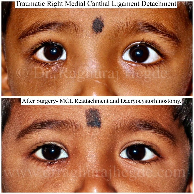
This child was mauled by a stray dog in the middle of the second wave of covid.  The child had injuries all around the face most prominently along the right lower eyelid, nose and a huge gash on the left cheek. 

The child was quickly taken up for primary suturing of facial wounds. During the wound exploration, we noted that the right MCL was ripped out off it’s bony attachment possibly along with the lacrimal sac. A very large hematoma was also noted in the same area which was cleared.  There was severe facial oedema during surgery so we chose to only do primary suturing of the eyelid and facial wounds at the time and to plan further management later. 

Post operatively on late review, the facial wounds had healed well but the child developed tearing (epiphora) on the right side and the child also had chronic dacryocystitis on the same side. The marked Telcanthus confirmed the MCL detachment noted intraoperatively. 

The CT scan images revealing a soft tissue swelling around right medial canthus including the lacrimal sac abscess (indicated by the red arrow)

A second surgery was performed. A  DCR was done using the pre-existing sub-ciliary scar continuing on to the upper lid. (See photo below to to see the marked U shaped incision). MCL was identified, scar tissue excised and MCL reattached to correct position. Distal canalicululi was stenosed and scarred. A DCR with bicanalicular silicone intubation was done. Mitomycin C was instilled locally to reduce fibroblastic activity and scarring. 

The outcome was extremely good as seen in the first picture. The child’s eyelid shape was restored, tearing and chronic dacryocystitis resolved. All this was done using aesthetically placed incisions recruiting the old scars from the initial injury. This has been an extremely satisfying surgery. 

Stay dogs can be an important cause of trauma in India. It is especially more common in small kids who are about the size of a grown up dog or smaller. It is advisable to keep kids away from exposure to stray dogs on the streets which can get suddenly violent if they perceive the kid as a threat. Dogs are great and cute creatures but can be dangerous to kids in certain circumstances. 

For Appointments:
📠 Phone: +91 80 2502 3257
📩 Email: dr.raghuraj.hegde@gmail.com
🖥️ Website: www.drraghurajhegde.com

©All patient photos are being used with the express consent of the patient. These cannot be shared or reproduced elsewhere. 

Dangers Lurking Around the Eye

This little 6yr old girl with recent history of URTI was quickly followed with eye swelling up. He photo shows her at presentation (Above) and after treatment -both surgical and medical treatment. (Below)

The CT scans which confirmed the clinical findings.

The dotted red circle showing a sub-periosteal abscess entering the orbit(eye socket). 

Hospital admission and IV antibiotics didn’t reduce the abscess and therefore we had to proceed with medial  orbitotomy via transcaruncular incision and drainage of abscess while the Rhinology team performed a limited Functional Endoscopic Sinus Surgery (FESS) to drain multiple sinuses full of mucopus. Below is the collage of the surgery. (viewer discretion advised)

Eye is surrounded by the Para-nasal sinuses. In children, the sinuses are small and they increase in size as the child grows. Children are vulnerable to sinus infections as a consequence of upper respiratory tract infections reducing the ciliary clearance from the sinuses and fluid stasis. Sinus infections in turn can be dangerous to the eyes as the distance between sinuses and eye is very small in children especially the medial walls of the orbits which is paper thin aptly named- lamina paprycea. 

Once the infection reaches the eye socket (orbit), it can quickly escalate to involving the orbital fat, various nerves and muscles within the eye socket including the optic nerve. Pus can collect in the form of an abscess adjacent to the eyeball (globe) push it forward. The threshold for intervention is very low in the case of small children. 

Where is the scar?

Try to spot the incision scar in the above photo. It’s there I assure you because an external Dacryocystorhinostomy (DCR) was done using an external skin incision. 

I always remember my mentor’s words while I was closing up during surgery during my fellowship,

“You may do the most complicated surgery in the most beautiful way possible but the patient will judge the surgery by the scar you leave behind”

This maxim has been drilled into me- so much so that my residents find me being too anal about wound closures. I spend extra time being careful about how I close wounds.

As oculoplastic surgeries move towards minimally invasive approaches with smaller and smaller incisions, the scar after surgery is something we need to discuss with patients. With the advent of Endoscopic Endonasal  Dacryocystorhinostomy (Endo-DCR) being as successful as external DCR, the absence of an external scar is an important selling point for Endoscopic surgeons.  

I offer both choices for patients but don’t oversell the scarless option of Endo-scopic DCR. Not discounting the Endoscopic skills required for Endo-DCR, it is possible to give good aesthetic outcome with external DCR and sometimes as in the case above, being as good as an Endo DCR!

✂️ Surgery : External Dacryocystorhinostomy
⏲Surgery time: 45 mins
💉Anaesthesia: Done under Local
🗓️Downtime: 2 weeks
📈Duration of effect: Long term 
📠 Phone: +91 80 2502 3257
📩 E-mail:dr.raghuraj.hegde@raghurajhegde
🖥️ Website: www.drraghurajhegde.com

©All patient photos are being used with the express consent of the patient. These cannot be shared or reproduced elsewhere. 

Your eyes reflects your personality

A young professional had recently joined the workforce. His clients, colleagues and seniors thought of him as a disinterested employee despite him working as hard as anyone else.

Why you ask?

He has had mild droopy eyelids (known as ptosis) since birth. It was not affecting his day to day functioning and vision when he is looking straight. However, the symmetrically droopy eyelids made it look like he was disinterested in the work he was doing- both when he was in college and now when he is working. 

This problem can be easily treated with a day care, single sitting surgery and there need not even be a external skin incision. The surgery is known as conjunctivo-mullerectomy where the ptosis (droopy eyelid) is corrected using an incision on the wet surface of the eyelid. The surgery and recovery is faster than conventional ptosis correction surgery. The outcomes of this surgery are very predictable if the patient selection is done correctly by the treating oculoplastic surgeon- as was in this case. 

This young man was thrilled with the final outcome of the surgery and he got to reclaim his real personality. 

⏲Surgery time: 20 mins
💉Anaesthesia: Done under Local
🗓️Downtime: 2 weeks
📈Duration of effect: Long term 
📠 Phone: +91 80 2502 3257
📩 E-mail:dr.raghuraj.hegde@gmail.com
🖥️ Website: www.drraghurajhegde.com

©All patient photos are being used with the express consent of the patient. These cannot be shared or reproduced elsewhere.