
Naso-Orbito-Ethmoidal (NOE) Fracture
I got a pleasant surprise a few days back when one of my patients who is now in another city sent me his selfie showing that he has got back to his old handsome self. He was operated by our team nearly a year back. I was very happy to know all our efforts was worthwhile.

Read on to know his surgical story.
This young man unfortunately got injured when he slipped while hurrying down the stairs and hit his face on the railing. He sustained what is known as Naso-Orbito-Ethmoidal (NOE) fracture. The frontal bone (forehead area) was severely fractured into multiple pieces- the force so high it caused leakage of his Cerebro-spinal fluid (fluid surrounding the brain). Both his orbits (Eye sockets) had large Blow-in fractures and his nose was flattened due to the force.
Our team consisting of a Neuro-surgeon, Oculoplastic Surgeon (Me) and Maxillofacial Surgeon operated on him for nearly 10 hours to get this young man back to his pre-accident status. The Neurosurgeon fixed his CSF leak after taking a bi-coronal flap approach and we then painstakingly fixed his frontal bone fractures piece by piece using titanium plates and mesh as a framework. I then repaired both his orbits👀 with pre-fabricated combined medial and floor titanium implants using the trans-conjunctival approach to avoid any extra skin incisions. The left anterior nasal buttress had to be fixed with a titanium mini-plate from an oral incision. The flattened nose 👃still had to be fixed which was then suspended from the frontal bone titanium frame while using internal nasal splints and external POP splint to hold the nasal bones in the desired position. This was perfect team work which reflected in the excellent post-operative outcome.
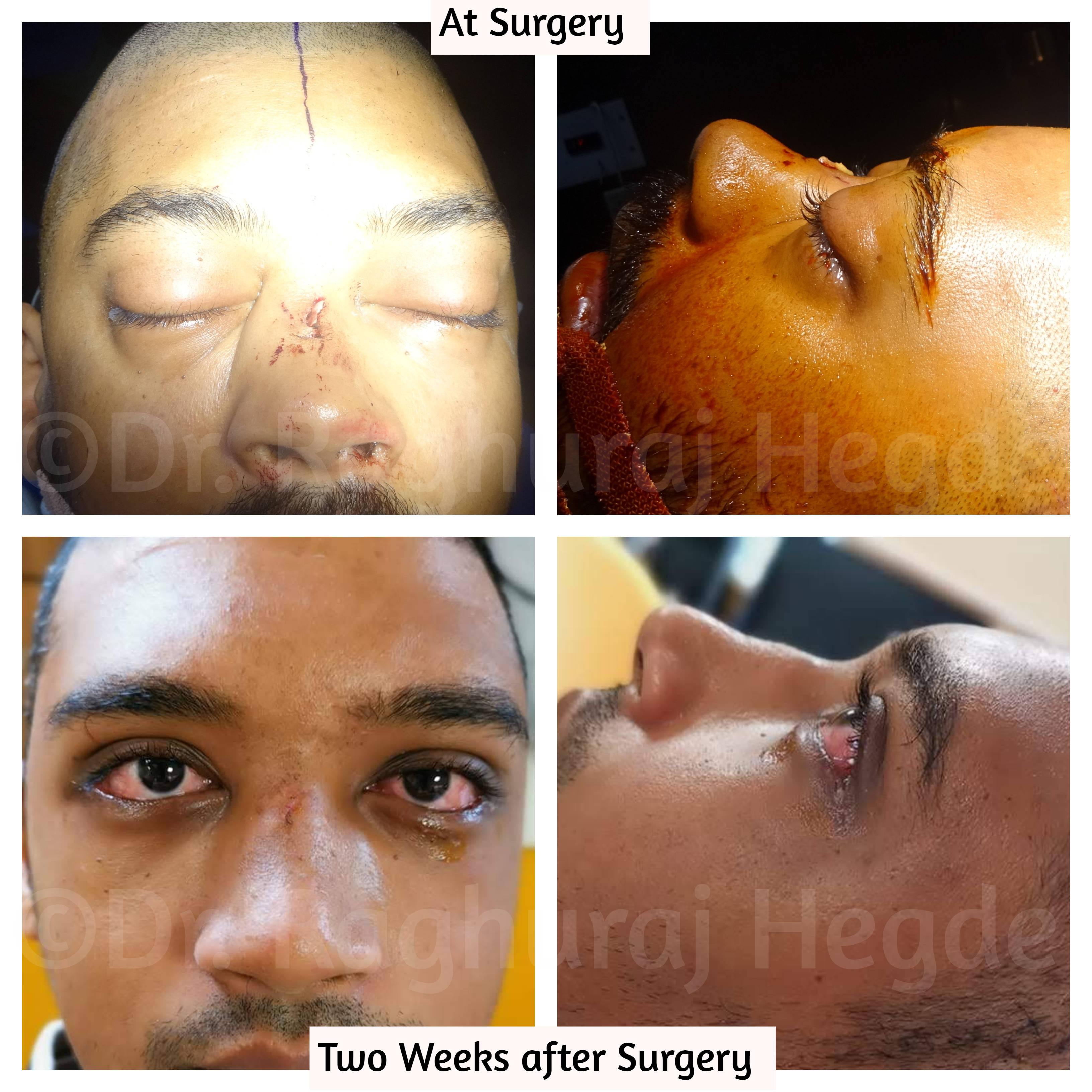
(Above) The patient’s Straight Face and Side Profile pictures on table and (Below) His Straight Face and Side Profile two weeks after surgery.
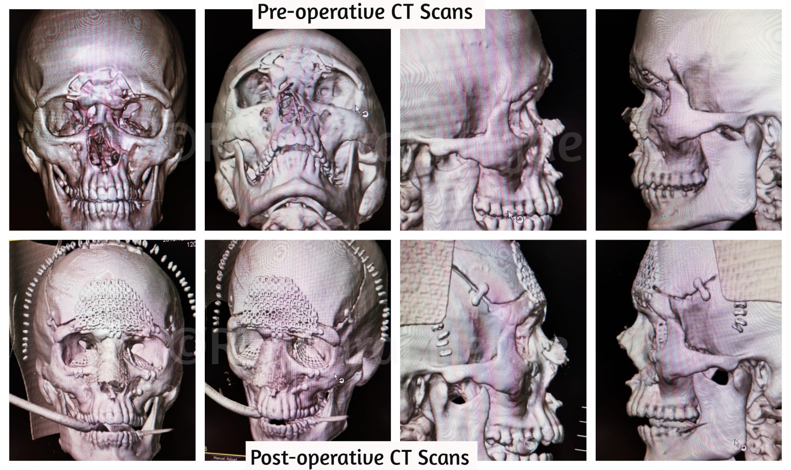
(Above) Pre-operative 3D reconstructed CT images showing severe frontal and nasal bone fractures. (Below) Post-operative 3D reconstructed CT images showing well corrected external fractures.

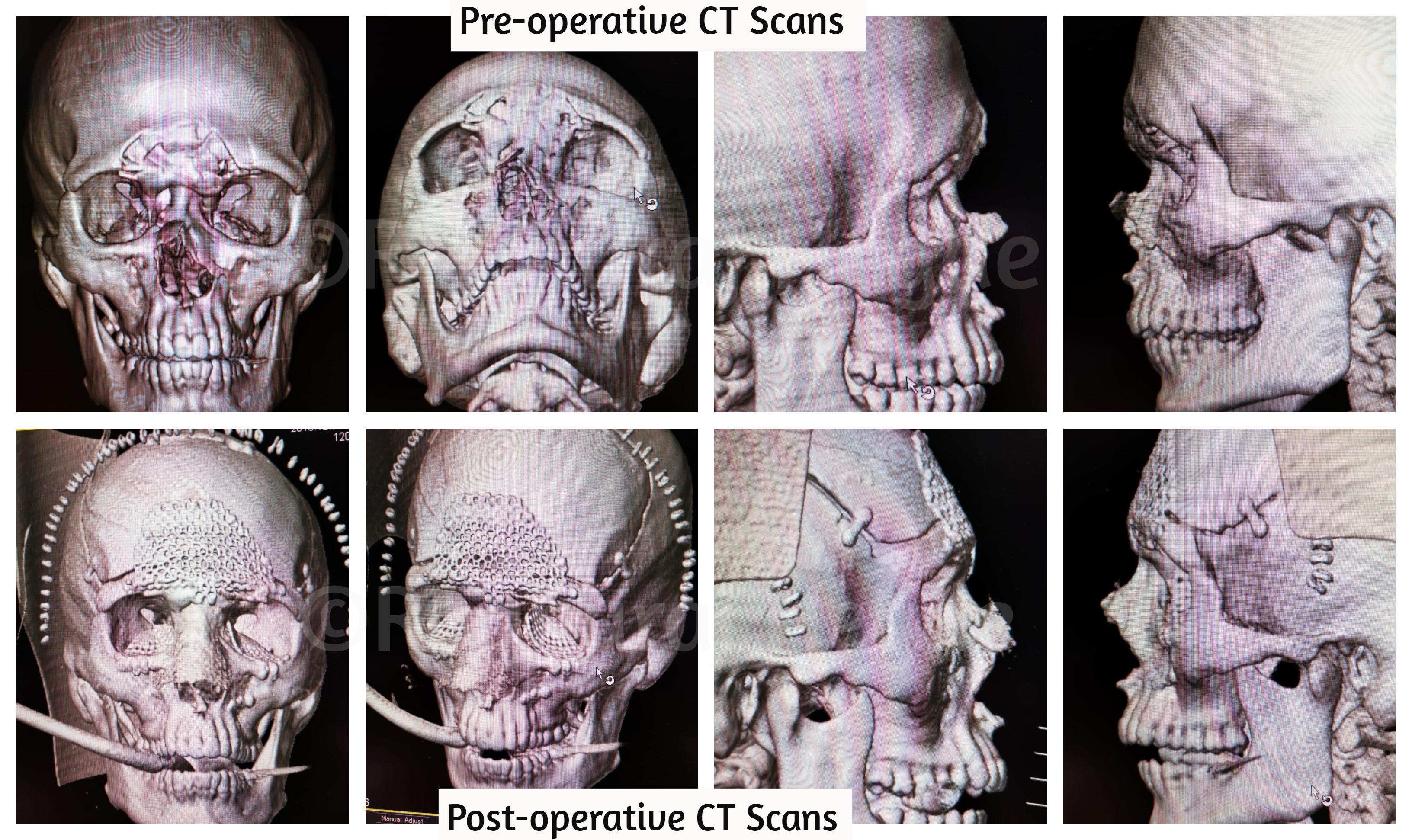
(Above) Pre-operative Coronal, Sagittal, Axial CT images showing Bilateral Blow-in floor and medial wall orbital fractures as a part of the NOE fracture. (Below) Post-operative Coronal, Sagittal, Axial CT images showing perfectly reconstructed orbits on both sides.
For details of such types of surgeries and for appointments
📠 Phone: +91 80 2502 3257
📩 E-mail:dr.raghuraj.hegde@gmail.com
🖥️ Website: www.drraghurajhegde.com
©All patient photos are being used with the express consent of the patient. These cannot be reused, shared or reproduced elsewhere without the consent of Dr. Raghuraj Hegde.
