

Once the incision wound had completely healed, it was like he never had a surgery. Even the patient himself couldn’t locate the scar. The is the benefit of a minimally invasive surgery and aesthetically planned surgery. Even though the entire orbital lobe of the lacrimal gland was excised, there was no dry eye in the patient. This is because most of the regular lacrimation is by minor lacrimal glands spread out throughout the conjunctiva (transparent layer on the eyeball).
Tag Archives: Orbit
Not all masses are tumours!

The histopathology of the mass turned out to be characteristic of tissue infected with Mycobacterium Tuberculosis. In the sub-continent, tuberculosis (TB) is always a differential even when tests for TB turn out to be negative. Histopathology is gold standard for diagnosing TB. Orbital TB though not always a top clinical diagnosis it is not uncommon.
Growing out of a Crack!

Dermoid cyst (also known as choristoma) is a benign tumor growing out of a embryonic suture line. The tumour consists of normal cells occurring in an abnormal location. It is usually diagnosed in children when it is first visible but it is not uncommon to have adults coming to the clinic to know if it can be removed.
Hugging the Optic Nerve!
The MRI scan showed a large intraconal well defined mass sitting right next to the optic nerve causing mass effect which in turn was causing diminution of vision. My working differentials going into surgery- based on location and imaging was Cavernous Hemangioma, Fibrous Histiocytoma, Dermoid cyst, Schwannoma among others.
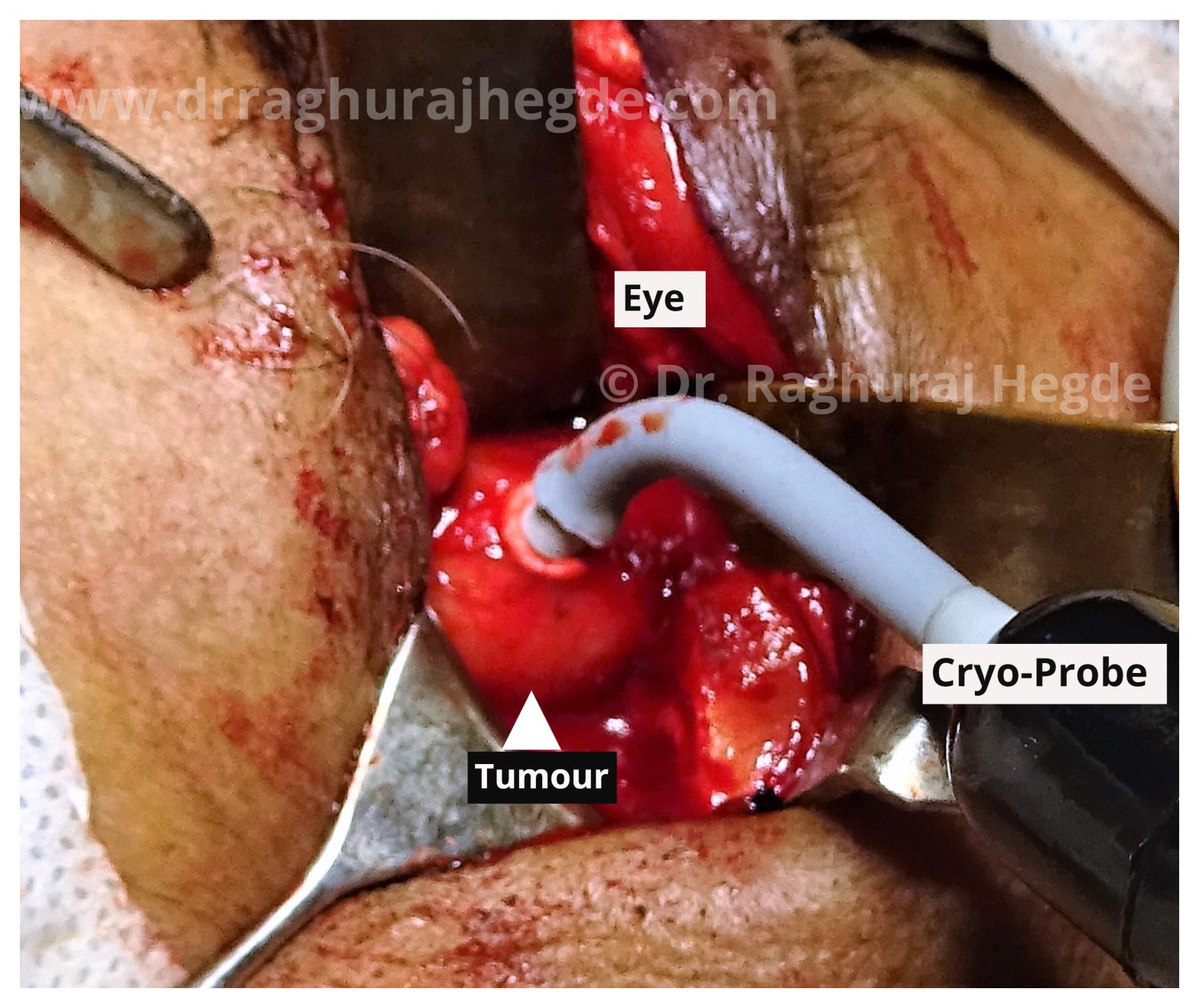
A Bridge between Eye & Brain
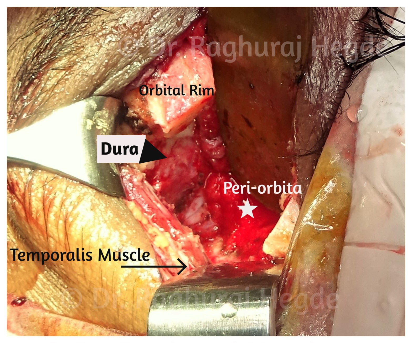
Nothing beats the feeling of being able to remove a skull base tumour through a small incision in the upper eyelid crease. This here is after the tumour has been completely excised. This picture shows the amount of exposure we can achieve by just the orbital route and also that’s me admiring the orbital anatomy for a few seconds before closing up.
A Nose to Remember!
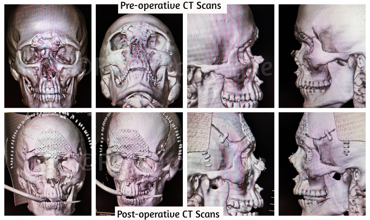
Our team consisting of a Neuro-surgeon, Oculoplastic Surgeon (Me) and Maxillofacial Surgeon operated on him for nearly 10 hours to get this young man back to his pre-accident status. The Neurosurgeon fixed his CSF leak after taking a bi-coronal flap approach and we then painstakingly fixed his frontal bone fractures piece by piece using titanium plates and mesh as a framework. I then repaired both his orbits👀 with pre-fabricated combined medial and floor titanium implants using the trans-conjunctival approach to avoid any extra skin incisions. The left anterior nasal buttress had to be fixed with a titanium mini-plate from an oral incision. The flattened nose 👃still had to be fixed which was then suspended from the frontal bone titanium frame while using internal nasal splints and external POP splint to hold the nasal bones in the desired position. This was perfect team work which reflected in the excellent post-operative outcome.
Finding a road to the fistula
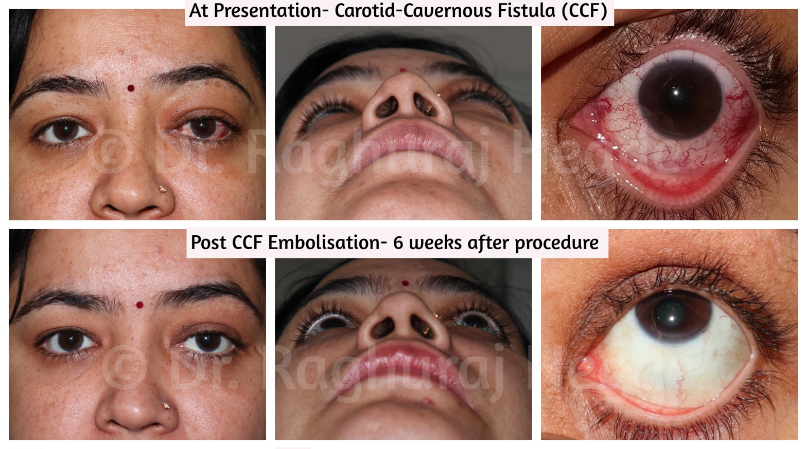
It is very rare that oculoplastic surgeons are asked to provide access to interventional neuro-radiologists. This is one such case. We have performed such interventions in 3 cases till date at our hospital with similarly successful outcomes. The important thing to remember as a surgeon while accessing the SOV is that the vein is arterialised due to the CCF and bleeding can be very severe is the vessel wall is damaged.
Blepharoplasty- Eyelid Lift Surgery
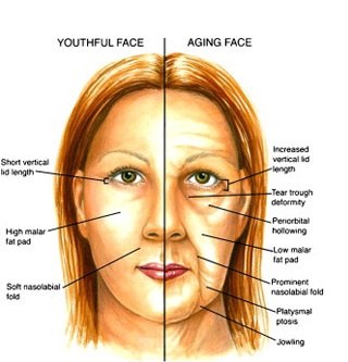
Blepharoplasty is the removal and repositioning of the skin, muscle and fat of the upper and/or lower eyelid. In the upper eyelid the incision is made and hidden in the natural lid crease. In the lower eyelid the incision is made along the skin just beneath the eyelashes or in the moist surface of the eyelid known as conjunctiva.
